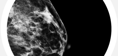
Screening Mammogram
Digital Screening Mammography is a safe, low-dose X-ray of the breast for women not experiencing any symptoms or other concerns about breast health. It is designed to identify cancer in the early stages, long before it can be detected by a physical examination. Screening Mammography typically consists of four views – two of each breast. It remains our best method of detecting breast cancer early.
Cypress Imaging is the region’s leading provider of digital mammography and offers the only facility in the region accredited by the American College of Radiology as a Breast Imaging Center of Excellence. At Cypress Imaging, all mammograms are done in a private and comfortable setting by specially trained, registered mammography technologists. These images are read by our experienced team of radiologists, and findings are sent to your primary care physician.
In addition to regular mammograms, we recommend monthly breast self-examination.
3D Mammography (Digital Breast Tomosynthesis)
3D Mammography is a game changer in breast cancer screening, detecting cancers earlier when they are easiest to treat.
- What is 3D Mammography?
3D Mammography, also known as breast tomosynthesis, converts images into thin layers building what is essentially a “Three Dimensional Mammogram”. Now, radiologists can see breast tissue in a way never before possible. With traditional mammography, radiologists view the complexities of the breast in a flat image. With 3D Mammography, breast tissue is viewed in thin, 1-millimeter slices. Fine details are clearly visible, no longer hidden by overlapping tissue. - What are the benefits?
3D Mammography allows our physicians to better see the different structures as well as the location, size and shape of any abnormal tissue. This allows more cancers to be found at earlier stages when they are more treatable and also reduces the chance of a false positive exam resulting in additional imaging. - What are the risks?
The amount of radiation is below the American College of Radiology guidelines and is equivalent to getting a digital 2D mammogram. Breast tomosynthesis is approved by the Food and Drug Administration (FDA). - The 3D Mammography exam
A tomosynthesis exam is very similar to a traditional 2D mammogram. Your breasts are under compression while the x-ray arm of the machine makes a quick arc over the breast, taking a series of images at a number of angles. All images are viewed by the technologist to ensure they have captured adequate images for review by our radiologist. The entire exam time should be approximately the same as that of a traditional mammogram. - Is there an additional cost?
Medicare covers the cost of 3D imaging. As always, please check with the insurance provider. If it is not covered, and you choose to have the additional 3D imaging, you will be asked to pay a minimal out of pocket fee.
CAD (Computer-Aided Detection)
In addition to initial review and interpretation of images by our radiologists, Mammograms at Cypress Imaging are reviewed using Computer Aided Detection (CAD) technology, which can highlight suspicious areas for additional evaluation.
Diagnostic Mammogram
When a Digital Screening Mammogram uncovers suspicious areas or a patient is having specific breast problems, a Diagnostic Mammogram is needed. This may consist of additional views beyond the standard four included in a Screening Mammogram. These additional views are also commonly used for women who have breast implants because the implants interfere with imaging the entire breast.
Ductogram
If a woman is experiencing drainage not associated with routine causes, a Ductogram may be performed. Ductography is a special type of contrast-enhanced mammography used for imaging the breast ducts. A Ductogram can aid in diagnosing the cause of an abnormal nipple discharge and is valuable in diagnosing intraductal papillomas and other conditions.
Scheduling Your Mammogram
It is best to schedule your mammogram the week after your period when the breast is less tender. Please let us know if you have breast implants so we can optimize your exam.
Preparing for Your Visit
If you have had mammograms done previously at other centers, arrange to have them sent to us prior to your visit, or bring copies with you. This will make it possible for our radiologist to compare and identify any changes that may be important, and ensure that you receive your results promptly without waiting for prior studies to arrive.
Do not wear deodorant, talcum powder, or lotion under your arms or on your breasts on the day of your visit. These products can often mimic breast disease.
During Your Visit
Before the exam, you will be asked to remove all jewelry and clothing from the waist up, and you will be asked to wear a gown that opens in the front
Please advise the technologist if you have any particular symptoms or can describe an abnormality. Also, be sure to mention any family history of breast cancer.
During your mammogram, the technologist will gently position each breast imaging plate to apply compression to the breast. Compression aids in visualizing the tissue and helps to see potential abnormalities that could be obscured by overlying breast tissue. You should expect to feel pressure from the compression, and some women who have sensitive breasts may feel discomfort. The compression will only last for a few moments and no discomfort should be felt following the exam. After each view, the breast will be repositioned to optimize any additional views.
After Your Visit
After your study is over, the images will be evaluated by one of our Board-Certified Radiologists who specialize in breast imaging. A final report will be sent to your physician who can then discuss the results, and any follow-up recommendations with you in detail.
Mammogram Callbacks
Learn what it really means when you’re called back for additional testing after your screening mammogram.
